Human Study Finds Early Visual Cortex Disinhibition Necessary for Long-Term Fear Memory
Nature Communications on November 14 published a human fear-memory study titled 'Sensory cortical disinhibition underlies long-term fear memory in humans'. How transient threats turn into persistent, long-term fear memories is a fundamental problem in neuroscience, and the underlying mechanisms had remained elusive.
The team combined high-definition α-frequency transcranial alternating current stimulation (HD α-tACS) with simultaneous EEG-fMRI and transformer-based neural decoding, tracking fear memory across formation, consolidation, and long-term retention, assessed at 15 minutes, 24 hours, and 7 days after fear conditioning.
Spatiotemporal memory traces were identified at all three assessments, beginning at an early sensory stage 40 to 60 milliseconds post-stimulus and distributed across visual cortex and limbic regions. Conditioning potentiated occipital α-oscillation desynchronization, a marker of visual cortical disinhibition, which subsequently predicted the strength of long-term memory traces.
Targeted α-tACS over early visual cortex induced early visual cortical (V1/V2) inhibition, abolished conditioning-potentiated α desynchronization, and selectively disrupted fear-memory consolidation and retention while leaving initial acquisition intact. The authors conclude that sensory cortical disinhibition is a necessary mechanism for long-term fear memory in humans and identify early sensory cortex as a crucial node for intervention in persistent maladaptive fear. The research was supported by National Institutes of Health grants R21MH126479 and R01MH132209, and the authors include Brown, Ma and Wang.
Source: Nature Communications
Published by GSM · USA (original Chinese edition)
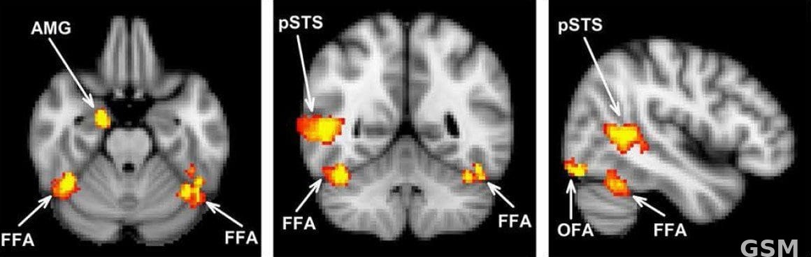
Photo: 图:功能性磁共振成像(fMRI)显示的面孔敏感脑区(资料图)。来源:Richard J. Harris、Andrew W. Young、Timothy J. Andrews,Wikimedia Commons,CC BY 3.0。 / Photo: Face-sensitive brain regions shown by functional MRI (file image). Source: Richard J. Harris, Andrew W. Young and Timothy J. Andrews, Wikimedia Commons, CC BY 3.0.