
UCLA-Led Team Maps Developing Human Basal Ganglia: 155,000 Cell Nuclei Traced Through Seven Months After Birth
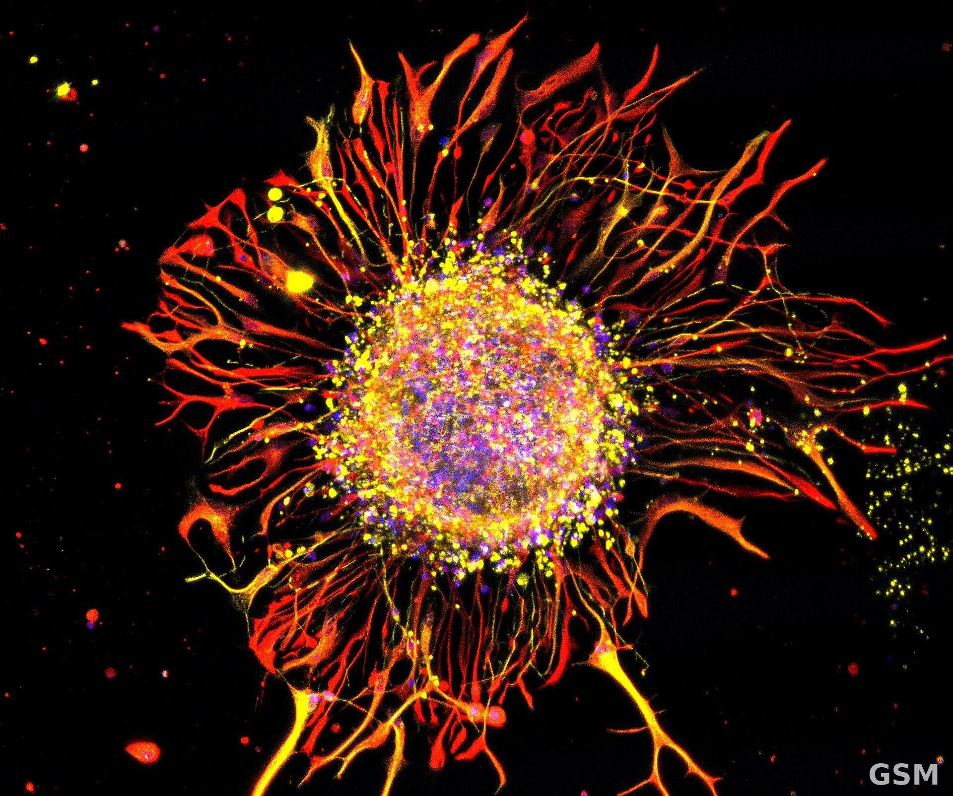
A research team co-led by UCLA on Nov. 16 released a detailed atlas of how cells in and around the basal ganglia develop before and after birth, in a study published in Cell. The basal ganglia are a group of structures deep in the brain that help control movement, habits, reward and behavior, and are involved in conditions such as Parkinson's disease, Huntington's disease, addiction and schizophrenia.
The research is part of the National Institutes of Health's BRAIN Initiative Cell Atlas Network (BICAN). Using a technology co-developed by the lab of co-senior author Chongyuan Luo, an associate professor of human genetics at UCLA's David Geffen School of Medicine, researchers analyzed over 155,000 cell nuclei from nine developing human brains, collecting data on DNA regulation from mid-pregnancy to seven months after birth. Combined with earlier data, the analysis covered nearly 287,000 cells.
The study showed that basal ganglia cells mature on different timelines from midpregnancy through infancy, undergoing major changes in how their DNA is regulated and organized. Some cells begin in temporary structures, the ganglionic eminences, that largely disappear around birth, and the team followed those cells from their starting points to where they eventually settle.
Comparing the atlas with genetic studies of psychiatric disorders, the researchers found that spiny projection neurons, a major type of neuron in the basal ganglia, showed especially strong overlap with genetic variants associated with schizophrenia. Luo said genetic studies can point to regions of the genome associated with disease but often cannot say which cell type is affected or when during development that genetic risk becomes relevant, and that this atlas provides that cellular and developmental context.
Related:
Researchers Map the Brain's Behavioral Gatekeeper for the First Time in NIH-Funded Atlas
(Source: UCLA)
Source: UCLA
Published by GSM · USA (original Chinese edition)
Photo: 图:神经元显微图像(Madalena Simões-Abade 摄)。经 Wikimedia Commons,CC BY-SA 4.0。 / Photo: Neurons under a microscope, by Madalena Simões-Abade. Via Wikimedia Commons, CC BY-SA 4.0.
 UCLA Puts a Lid on Cell-Scale Test Tubes: Capped Nanovials Hold Bacteria and Yeast and Let Cells Grow Into Colonies2026-11-16
 Scientific Reports Paper on November 16: AI-Assisted Ultrasonography Detects Moderate-to-Severe Kidney Interstitial Fibrosis With Pooled Sensitivity of 0.821 and Specificity of 0.828 Across 14 Studies and 3,776 Participants2026-11-16